Key takeaways
- Biological Asymmetry: The human body regulates iron absorption via the hormone hepcidin, but lacks any active excretion pathway — men and postmenopausal women steadily accumulate iron stores over decades.
- Fenton Reaction & Oxidative Stress: Poorly bound and free ferrous iron (Fe2+) reacts with hydrogen peroxide (H2O2) to generate hydroxyl radicals (OH•), initiating lipid peroxidation, damaging mitochondrial DNA, and promoting ferroptosis.
- Longevity vs. Conventional Ranges: While hospital reference intervals often accept ferritin up to 300–400 µg/L as 'normal', longevity and epidemiological evidence indicates that the optimal window for healthy aging spans 40 to 120 µg/L.
- Dual-Biomarker Necessity: Elevated ferritin (>200 µg/L) must always be evaluated alongside transferrin saturation (TSAT) and hs-CRP; high ferritin with normal TSAT frequently reflects metabolic inflammation (MASLD), whereas high TSAT (>45%) indicates authentic systemic iron accumulation.
- Proven Intervention: A single standard whole blood donation (450 ml) removes approximately 200–250 mg of elemental iron, typically reducing serum ferritin by 30–50 µg/L, making routine blood donation one of the most potent longevity interventions for men.
Medical disclaimer: Content is for informational purposes and does not replace medical advice.
The Biology of Ferritin: From Vital Storage to Fenton Reaction Free Radicals
Iron is a quintessential double-edged sword in human biology. On one side, this transition metal is utterly essential: it binds oxygen in hemoglobin, stores oxygen in myoglobin within skeletal muscle, and serves as an indispensable catalytic cofactor for electron transport chain cytochromes within mitochondria, where cellular ATP is generated. Without adequate iron, cellular respiration collapses, producing profound physical exhaustion, pallor, and cognitive decline. PMID 11268303 PMID 24795252
On the other hand, unbound free iron is chemically unstable and highly cytotoxic. To prevent free iron ions from circulating unprotected through the bloodstream and delicate tissues, evolution engineered specialized transport proteins (transferrin) and intracellular storage nanocages (ferritin). Ferritin is a spherical protein shell composed of 24 subunits (heavy H-chains and light L-chains) capable of sequestering up to 4,500 iron atoms in a non-reactive, crystalline ferric core (Fe3+). Under steady-state physiological conditions, circulating serum ferritin correlates closely with total body iron reserves. PMID 11268303 PMID 24795252
The pathological cascade begins when storage capacity is overwhelmed or when local microenvironments turn acidic and pro-inflammatory. Under these conditions, ferrous iron (Fe2+) dissociates, sparking the classical Fenton reaction: PMID 11268303 PMID 24795252
Fe²⁺ + H₂O₂ → Fe³⁺ + OH• + OH⁻ PMID 11268303 PMID 24795252
Through this chemical transformation, relatively benign hydrogen peroxide (H2O2), produced naturally during oxidative phosphorylation, is converted into the extraordinarily destructive hydroxyl radical (OH•). Hydroxyl radicals possess a half-life measured in nanoseconds and instantaneously react with whatever biological macromolecule they strike. They trigger chain reactions of lipid peroxidation within cell membranes, oxidize endothelial vascular surfaces, accelerate oxidized LDL (oxLDL) formation, and inflict direct double-strand breaks on mitochondrial and genomic DNA. PMID 11268303 PMID 24795252
In recent years, molecular geroscience has highlighted ferroptosis — an iron-dependent, non-apoptotic form of regulated cell death driven by toxic accumulation of lipid hydroperoxides. Chronic tissue iron excess primes cardiomyocytes, neurons, and hepatocytes for ferroptotic degeneration, directly driving accelerated biological aging. PMID 11268303 PMID 24795252
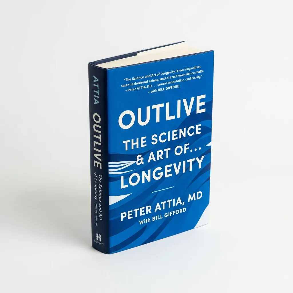
Outlive: The Science & Art of Longevity
Dr. Peter Attia's groundbreaking guide to preventive medicine, exercise for longevity, and healthy aging.
- Scientific framework for Medicine 3.0
- Actionable guide on sleep, exercise & nutrition
Reference & Risk Matrix: Conventional Ranges vs. Longevity-Optimal Targets
A major challenge in contemporary preventive healthcare stems from the broad reference intervals utilized by standard clinical laboratories. Hospital lab reports routinely classify serum ferritin levels between 30 and 400 µg/L in adult men (and 15 to 200–300 µg/L in postmenopausal women) as entirely 'normal'. These wide brackets are based on statistical population distributions in cohorts that frequently harbor subclinical steatotic liver disease, metabolic syndrome, and unmanaged iron accumulation, rather than biomarker levels that optimize disease-free longevity. PMID 24795252 PMID 20427843
Longevity physicians and hematologists recommend a significantly tighter target window. The matrix below contrasts conventional clinical benchmarks against optimal longevity targets and outlines actionable clinical steps based on transferrin saturation (TSAT). PMID 24795252 PMID 20427843
| Serum Ferritin (µg/L) | Clinical Status | Longevity Assessment | Transferrin Saturation (TSAT) | Biochemical Mechanism & Action |
|---|---|---|---|---|
| < 20–30 µg/L | Iron Deficiency / Anemia Risk | Suboptimal (Cellular Hypoxia) | < 16–20 % | Impaired cytochrome and ATP synthesis. Verify hemoglobin; evaluate iron bisglycinate supplementation under medical guidance. |
| 40–120 µg/L | Conventional 'Normal' | Gold Standard / Longevity Optimal | 20–35 % | Optimal equilibrium: adequate iron for mitochondrial respiration and erythropoiesis without catalytic Fenton reaction risks. |
| 120–200 µg/L | Conventional 'Normal' | Mild Accumulation (Acceptable) | 30–40 % | Moderate stores. For healthy adult men and postmenopausal women, an ideal threshold to initiate annual or bi-annual blood donation. |
| 200–350 µg/L | High Normal / Gray Zone | Elevated Oxidative Risk | 35–45 % (or normal TSAT in MASLD) | Assess hs-CRP and ALT. If hs-CRP is low and TSAT exceeds 40%, actively deplete excess iron stores through therapeutic phlebotomy. |
| 350–600 µg/L | Clinically Elevated | Substantial Pro-Oxidative Burden | > 45–55 % | Accelerated endothelial damage and hepatic stress. Screen for HFE gene mutations (hemochromatosis) and initiate structured phlebotomy. |
| > 600–1,000+ µg/L | Pathologically High | Toxic / Organ-Damaging Burden | > 50–80 % | Imminent risk of hemosiderosis, liver fibrosis, cardiomyopathy, and bronze diabetes. Requires urgent specialist hematological care. |
Differential Diagnosis: Acute-Phase Reactant (Inflammation) vs. True Iron Overload
A common diagnostic pitfall in preventive medicine is assuming that every elevated ferritin measurement reflects bona fide iron overload. Ferritin functions as a prominent positive acute-phase reactant. Hepatocytes in the liver and reticuloendothelial macrophages upregulate ferritin synthesis and secretion in response to inflammatory cytokines, particularly interleukin-6 (IL-6) and tumor necrosis factor-alpha (TNF-alpha). Evolutionarily, this sequesters free iron to starve invading pathogens that rely on host iron for replication. PMID 24795252 PMID 33130635
When a lab result returns with serum ferritin at 320 µg/L, two radically different physiological realities are possible: PMID 24795252 PMID 33130635
**Scenario 1: Metabolic Inflammation and Steatotic Liver (MASLD / NAFLD)**: If the individual presents with mildly elevated high-sensitivity C-reactive protein (hs-CRP > 1.5–2.0 mg/L), elevated alanine aminotransferase (ALT > 35–40 U/L), or homeostatic model assessment of insulin resistance (HOMA-IR > 2.0), while transferrin saturation remains entirely normal (e.g., 24–32%), the elevated ferritin is primarily a marker of systemic metabolic inflammation and hepatic stress. The patient does not necessarily require iron unloading; the clinical priority is reversing visceral adiposity and insulin resistance. PMID 24795252 PMID 33130635
**Scenario 2: True Biological Iron Overload**: If ferritin is 320 µg/L, hs-CRP is undetectable (< 0.8 mg/L), and transferrin saturation exceeds 45–50%, the elevation reflects authentic saturation of the body's iron-binding capacity. Free and labile iron pools are expanded. This scenario is frequently observed in middle-aged men with high red meat intake who do not donate blood, or in carriers of hemochromatosis variants. PMID 24795252 PMID 33130635
To achieve clinical precision, an iron assessment should never consist of an isolated ferritin test, but must include a complete iron panel: serum iron, transferrin, transferrin saturation (TSAT), complete blood count (hemoglobin), ALT, and hs-CRP. PMID 24795252 PMID 33130635
Why Men and Postmenopausal Women Accumulate Iron: HFE Mutations and the Multivitamin Error
From an evolutionary standpoint, iron deficiency was a perpetual threat to our ancestral forebears due to physical trauma, parasitic infestation, and scarce nutritional availability. Consequently, human physiology evolved robust mechanisms to absorb and conserve iron via hepatic hepcidin signaling, but zero active excretion pathways beyond minuscule daily sloughing of intestinal and epidermal cells (approximately 1 mg daily). PMID 20427843 PMID 30521876
Men do not experience cyclical blood loss. From the moment skeletal growth ceases in early adulthood, men begin accumulating a net surplus of 1–2 mg of dietary iron every day if consuming a standard Western omnivorous diet. By their fourth and fifth decades, typical men harbor three to five times more total body iron than age-matched women. PMID 20427843 PMID 30521876
In contrast, women remain naturally shielded from iron overload throughout their reproductive years by monthly menstruation, losing 15 to 40 mg of elemental iron per cycle. Many cardiovascular researchers view this physiological variance as the foundation of 'The Iron Hypothesis': premenopausal women's lower incidence of early coronary artery disease and four-to-five-year longevity advantage over men is partly attributed to their lower lifetime iron burden and subdued oxidative stress. However, once women transition through menopause, regular blood loss ceases. Within 5 to 10 years, postmenopausal women's ferritin levels climb rapidly, matching men's concentrations and precipitating an escalation in cardiovascular risk. PMID 20427843 PMID 30521876
**The HFE Gene and Hereditary Hemochromatosis**: PMID 20427843 PMID 30521876
Northern European populations exhibit a notably high prevalence of mutations in the homeostatic iron regulator (HFE) gene (predominantly C282Y and H63D). Approximately 1 in 8 individuals of Northern European descent carries at least one variant allele, and roughly 1 in 250 is homozygous (hereditary hemochromatosis). In affected individuals, the liver produces insufficient hepcidin, allowing the small intestine to absorb two to four times more dietary iron than physiological homeostasis requires. Without early detection and phlebotomy, this leads to iron accumulation across the liver, pancreas (bronze diabetes), joints, and myocardium. PMID 20427843 PMID 30521876
**The Critical Multivitamin Mistake**: PMID 20427843 PMID 30521876
One of the most counterproductive supplement errors occurs when men or postmenopausal women take generic daily multivitamins containing 10–14 mg of supplemental iron. Unless a physician has documented genuine microcytic iron deficiency anemia, men and postmenopausal women should never consume supplemental iron. Always examine product labels carefully and choose specialized iron-free multivitamin formulas (typically designated as 50+ formulations). PMID 20427843 PMID 30521876
Action Protocol: Practical Strategies to Lower Ferritin (Phlebotomy, Diet, and Scandinavian Realities)
If comprehensive blood testing reveals serum ferritin above 150–200 µg/L alongside transferrin saturation exceeding 35%, iron overload represents one of the most straightforward risk factors to correct in clinical precision medicine. The three-step protocol below outlines the evidence-based approach: PMID 30521876 PMID 33130635
**1. Voluntary Blood Donation as a Therapeutic Intervention**: PMID 30521876 PMID 33130635
The single most potent, physiological, and well-researched method to eliminate elemental iron is therapeutic phlebotomy. Each standard whole blood donation (450 ml) removes approximately 200–250 mg of elemental iron bound within erythrocyte hemoglobin. To regenerate the cleared red blood cells, the bone marrow mobilizes iron from intracellular ferritin stores in the liver and spleen. This routinely produces a predictable 30–50 µg/L reduction in serum ferritin per donation. PMID 30521876 PMID 33130635
For healthy adult men and postmenopausal women meeting local donor criteria (such as GivBlod in Denmark or national Red Cross blood banks across Europe), voluntary blood donation two to three times per year is not merely an altruistic civic contribution; it constitutes a direct longevity therapy. Landmark prospective cohort studies (including the Kuopio Ischemic Heart Disease Risk Factor Study) demonstrate that regular blood donors experience significantly lower rates of acute myocardial infarction and improved insulin sensitivity compared to non-donors. PMID 30521876 PMID 33130635
Please note: For individuals formally diagnosed with hereditary hemochromatosis via genetic testing and presenting with ferritin exceeding 500 µg/L, treatment is conducted as medical phlebotomy under hospital hematological supervision. PMID 30521876 PMID 33130635
**2. Nutritional Strategies and Absorption Inhibitors**: PMID 30521876 PMID 33130635
It is rarely necessary to transition to an extreme vegan diet, but targeted meal adjustments can substantially attenuate intestinal iron uptake: PMID 30521876 PMID 33130635
• **Polyphenols with meals**: Black and green tea contain tannins, and coffee contains chlorogenic acid, which chelate non-heme iron within the intestinal lumen and blunt absorption by 60–80%. Enjoy a cup of green tea or coffee alongside iron-dense meals. PMID 30521876 PMID 33130635
• **Calcium as a competitive inhibitor**: Calcium competes with iron for the divalent metal transporter 1 (DMT1) in enterocytes. Consuming dairy products or calcium-rich foods alongside meals inhibits both heme and non-heme iron uptake. PMID 30521876 PMID 33130635
• **Avoid high-dose vitamin C with iron-rich foods**: Ascorbic acid reduces insoluble ferric iron (Fe3+) to soluble ferrous iron (Fe2+), boosting absorption by up to 300%. Avoid high-dose vitamin C supplements around meal times containing red meat. PMID 30521876 PMID 33130635
• **Moderate red meat and offal intake**: Organ meats and red meat deliver readily bioavailable heme iron that bypasses natural mucosal block mechanisms. Restrict red meat to one or two meals weekly if stored iron is high. PMID 30521876 PMID 33130635
**3. Testing and Monitoring Cadence**: PMID 30521876 PMID 33130635
Serum iron biomarkers should be assessed via fasting morning blood panels, as circulating iron undergoes marked circadian fluctuations. After initiating regular blood donations, re-test ferritin and transferrin saturation after 3 to 6 months to ensure you do not overshoot into functional iron deficiency. Aim to stabilize your ferritin within the longevity sweet spot of 40 to 120 µg/L. PMID 30521876 PMID 33130635
Internal Further Reading
Read also in the same cluster
Magnesium Glycinate 400mg Capsules
Highly bioavailable chelated magnesium bound to glycine for maximum sleep and muscle absorption.
- Activates central GABA receptors for deep sleep
- Gentle on the stomach with superior bioavailability
FAQ
Why do standard blood tests often only measure ferritin and omit the full iron panel?
In primary care medicine, the prevailing goal is typically ruling out classic iron deficiency anemia, for which low ferritin (< 20–30 µg/L) is a sensitive standalone marker. However, when evaluating values above 150 µg/L, ferritin alone is insufficient because it non-specifically elevates during infection, systemic inflammation, or fatty liver. Differentiating true iron overload from inflammatory signaling requires measuring transferrin saturation (TSAT) and hs-CRP.
Can I lower high ferritin stores solely through dietary changes without blood donation?
Significantly reducing established high ferritin stores through diet alone is exceedingly slow. Because the human body sheds only about 1 mg of elemental iron daily through natural cell exfoliation, depleting an excess of 150 µg/L ferritin via dietary restriction would require multiple years. Dietary modifications are excellent for preventing future accumulation, but blood donation eliminates 200–250 mg of iron in a single 10-minute session.
What happens if my ferritin drops below 30 µg/L from donating blood too frequently?
If serum ferritin drops below 30–40 µg/L, you enter functional iron deficiency. Common symptoms include unexplained fatigue, diminished exercise capacity, lightheadedness, thinning hair, and impaired cognitive focus. Blood collection centers routinely check fingerstick hemoglobin, but do not always monitor ferritin at every visit. Pause blood donations if your ferritin approaches 40–50 µg/L.
Is elevated ferritin a verified risk factor for Alzheimer's and Parkinson's disease?
Yes, expanding neurogerontological research indicates that unmanaged systemic iron progressively accumulates within specific brain nuclei (including the substantia nigra and basal ganglia) with advancing age. Excess localized iron catalyzes the Fenton reaction, inflicting oxidative damage on oligodendrocytes and accelerating the aggregation of beta-amyloid and alpha-synuclein proteins.
Sources and References
- [1]
- [2]
- [3]
- [4]
Show all 5 sources (1 more)
- [5]
Editorial History
28. september 2026
Første publicering
Første version blev publiceret som del af precision medicine med intro, takeaways, FAQ og referenceblok.
28. september 2026
Faglig gennemgang
Formuleringer, forbehold og interne links blev gennemgået for klarhed, konsistens og YMYL-tydelighed.
28. september 2026
Seneste opdatering
Ferritin After 50 fik opdaterede metadata, referenceoutput og forbedret beslutningsnær struktur.

Medical Review
Medical Review and Update Process
This article has been medically reviewed by Vadim Doroshenko with a focus on clinical accuracy, sources, disclaimers, and whether the conclusions still align with current scientific evidence.

Editorial Profile
Borys Viktorovytj Riabtsev
Borys Viktorovytj Riabtsev publishes Longevity Lab as a research-backed information project on precision medicine, metabolic health, and healthy aging, utilizing rigorous sources, independent medical review, and objective YMYL communication.